|
GenScript corporation
girk4 cdna Girk4 Cdna, supplied by GenScript corporation, used in various techniques. Bioz Stars score: 90/100, based on 1 PubMed citations. ZERO BIAS - scores, article reviews, protocol conditions and more https://www.bioz.com/product/full+length+girk4+cdna/coding+dna+sequence+for+human+girk4+with+a+single+c+myc+tag/pm37549917-42-26-30 Average 90 stars, based on 1 article reviews
girk4 cdna - by Bioz Stars,
2026-11
90/100 stars
|
Buy from Supplier |
|
GenScript corporation
girk1 (kir3.1) girk4 (kir3.4) cdna Girk1 (Kir3.1) Girk4 (Kir3.4) Cdna, supplied by GenScript corporation, used in various techniques. Bioz Stars score: 90/100, based on 1 PubMed citations. ZERO BIAS - scores, article reviews, protocol conditions and more https://www.bioz.com/product/full+length+girk4+cdna/g+protein+coupled+inwardly+rectifying+potassium+channel++girk++1/pmc07577048-24-3-5 Average 90 stars, based on 1 article reviews
girk1 (kir3.1) girk4 (kir3.4) cdna - by Bioz Stars,
2026-11
90/100 stars
|
Buy from Supplier |
|
OriGene
molecular cloning human kcnj5 Molecular Cloning Human Kcnj5, supplied by OriGene, used in various techniques. Bioz Stars score: 90/100, based on 1 PubMed citations. ZERO BIAS - scores, article reviews, protocol conditions and more https://www.bioz.com/product/full+length+girk4+cdna/KCNJ5+(NM_000890)+Human+Untagged+Clone/us09982026-917-0-7 Average 90 stars, based on 1 article reviews
molecular cloning human kcnj5 - by Bioz Stars,
2026-11
90/100 stars
|
Buy from Supplier |
|
Alomone Labs
anti girk4 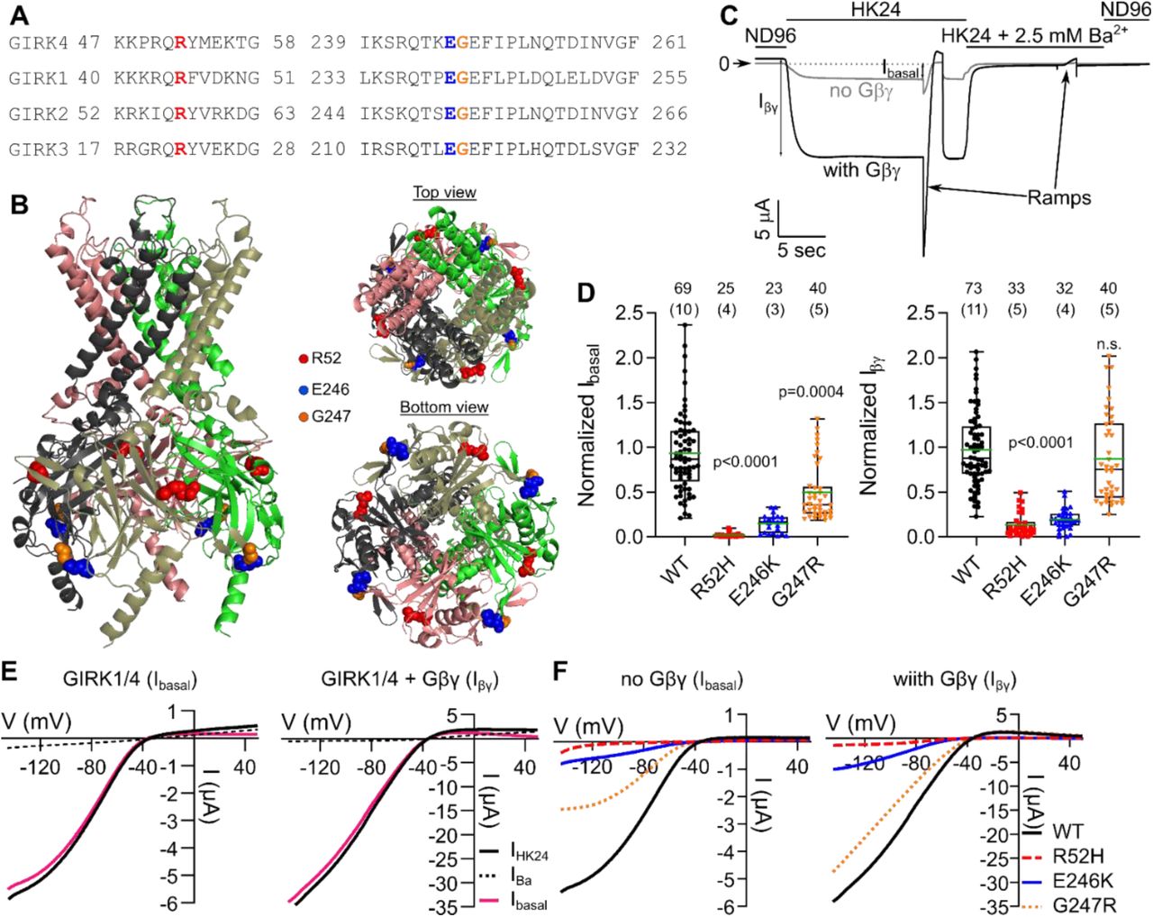 Anti Girk4, supplied by Alomone Labs, used in various techniques. Bioz Stars score: 95/100, based on 1 PubMed citations. ZERO BIAS - scores, article reviews, protocol conditions and more https://www.bioz.com/product/full+length+girk4+cdna/Anti-KCNJ5+(Kir3%2E4)+Antibody/bio_rxiv__866202-209-27-31 Average 95 stars, based on 1 article reviews
anti girk4 - by Bioz Stars,
2026-11
95/100 stars
|
Buy from Supplier |
|
Alomone Labs
tertiapin lq  Tertiapin Lq, supplied by Alomone Labs, used in various techniques. Bioz Stars score: 90/100, based on 1 PubMed citations. ZERO BIAS - scores, article reviews, protocol conditions and more https://www.bioz.com/product/full+length+girk4+cdna/Tertiapin-LQ/pmc09581561-74-5-15 Average 90 stars, based on 1 article reviews
tertiapin lq - by Bioz Stars,
2026-11
90/100 stars
|
Buy from Supplier |
Image Search Results
Journal: bioRxiv
Article Title: A revised mechanism of action of hyperaldosteronism-linked mutations in cytosolic domains of GIRK4 (KCNJ5)
doi: 10.1101/866202
Figure Lengend Snippet: (A) Alignment of GIRK4 with GIRK1, GIRK2, and GIRK3 shows that R52, E246, and G247R are conserved amino acids. (B) Crystal structure of GIRK2 marked with highlighted positions of the mutations; R52 – red, E246 – blue, and G247 – orange (numbering corresponds to GIRK4). (C) Example of the recording protocol in GIRK1/4 WT injected oocytes without (gray) or with (black) coinjected Gβγ. Horizontal bars show the corresponding external solutions. Voltage ramps were applied in HK24 and HK24 + 2.5 mM Ba 2+ solutions. (D) Normalized I basal (left) and I βγ (right). Currents were measured at V m =-80 mV. (E) Representative I-V relationships of currents of GIRK1/4 (I basal ; left) and GIRK1/4 + Gβγ (Iβγ ; right). Net GIRK currents were obtained by subtraction of I-V curve in Ba 2+ (“Ba 2+ -subtraction” procedure). (F) Representative net I-V relationships of GIRK1/4 and mutants; I basal (left) and I βγ (right).
Article Snippet: Protein samples (35µg) were electrophoresed on 12% polyacrylamide-SDS gel and transferred to nitrocellulose membranes for Western blotting with antibodies; anti-GIRK1 at 1:300 dilution (Alomone Labs, APC-005) or
Techniques: Injection

Journal: bioRxiv
Article Title: A revised mechanism of action of hyperaldosteronism-linked mutations in cytosolic domains of GIRK4 (KCNJ5)
doi: 10.1101/866202
Figure Lengend Snippet: A separate experiment was performed for each mutant. (A) Representative confocal images of YFP-GIRK4 expressing oocytes, with or without Gβγ. (B) YFP-GIRK4 expression in the PM; except for YFP-GIRK4 G247R +Gβγ, all mutants express much less than YFP-GIRK4 WT . (C) I βγ of YFP-GIRK4 homotetramers. All mutants were not active as homotetramers. (D) Representative I-V relationships of YFP-GIRK4 (WT and mutants) coexpressed with Gβγ.
Article Snippet: Protein samples (35µg) were electrophoresed on 12% polyacrylamide-SDS gel and transferred to nitrocellulose membranes for Western blotting with antibodies; anti-GIRK1 at 1:300 dilution (Alomone Labs, APC-005) or
Techniques: Mutagenesis, Expressing

Journal: bioRxiv
Article Title: A revised mechanism of action of hyperaldosteronism-linked mutations in cytosolic domains of GIRK4 (KCNJ5)
doi: 10.1101/866202
Figure Lengend Snippet: (A) Representative confocal images of GIRK1/YFP-GIRK4 expressing oocytes, with or without Gβγ. (B) GIRK1/YFP-GIRK4 expression in PM; mutated channels express less well than GIRK1/YFP-GIRK4 WT . A separate experiment was performed for each mutant. (C) Currents of GIRK1/YFP-GIRK4 WT and mutated channels. (D) Raw data from FRET experiments, showing E app as a function of donor/acceptor molar ratio. Each symbol corresponds to E app from one oocyte, from 4 experiments. (E) Binned analysis of E app shows that, at all donor/acceptor molar ratios, the interaction of CFP-Gβγ with GIRK1/YFP-GIRK4 R52H and GIRK1/YFP-GIRK4 E246K was impaired, whereas the interaction of CFP-Gβγ with GIRK1/YFP-GIRK4 G247R was more similar to GIRK1/YFP-GIRK4 WT . Molar ratios >2.5 were not included in the analysis because of insufficient data points forGIRK1/YFP-GIRK4 WT .
Article Snippet: Protein samples (35µg) were electrophoresed on 12% polyacrylamide-SDS gel and transferred to nitrocellulose membranes for Western blotting with antibodies; anti-GIRK1 at 1:300 dilution (Alomone Labs, APC-005) or
Techniques: Expressing, Mutagenesis
Journal: bioRxiv
Article Title: A revised mechanism of action of hyperaldosteronism-linked mutations in cytosolic domains of GIRK4 (KCNJ5)
doi: 10.1101/866202
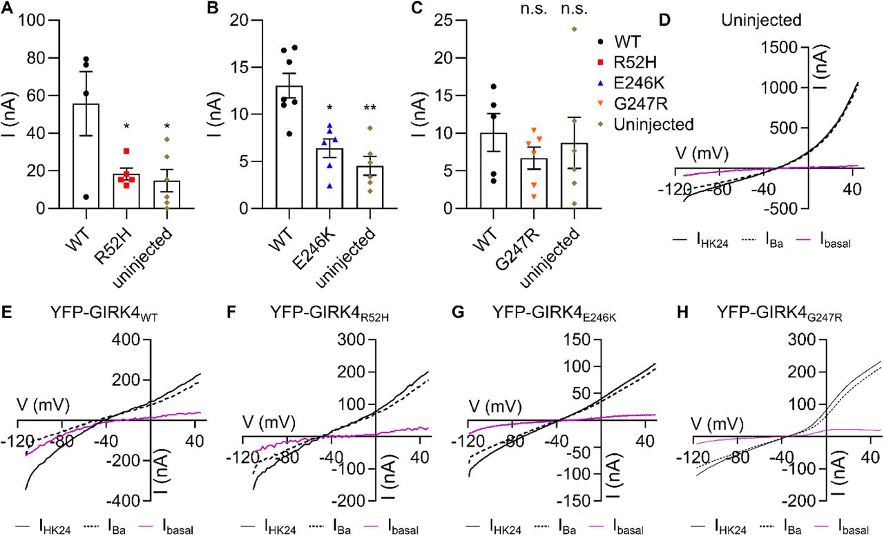
Figure Lengend Snippet: (A-C) Currents in oocytes injected with RNAs of YFP-GIRK4 mutants, measured at −80 mV, were indistinguishable from currents recorded in uninjected oocytes. (D-H) Representative I-V relationships of uninjected oocytes (D) and homotetrameric YFP-GIRK4WT (E), YFP-GIRK4R52H (F), YFP-GIRK4E246K (G), YFP-GIRK4G247R (H).
Article Snippet: Protein samples (35µg) were electrophoresed on 12% polyacrylamide-SDS gel and transferred to nitrocellulose membranes for Western blotting with antibodies; anti-GIRK1 at 1:300 dilution (Alomone Labs, APC-005) or
Techniques: Injection
Journal: bioRxiv
Article Title: A revised mechanism of action of hyperaldosteronism-linked mutations in cytosolic domains of GIRK4 (KCNJ5)
doi: 10.1101/866202
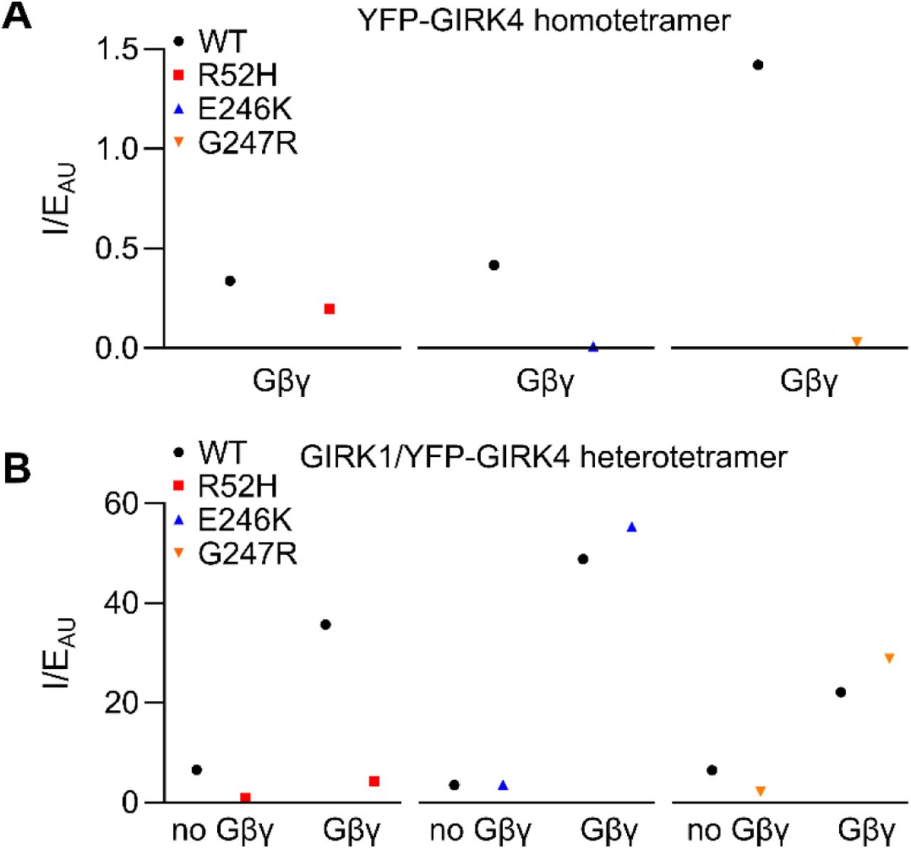
Figure Lengend Snippet: (A) Normalization of current to expression of YFP-GIRK4 homotetramers indicates an impaired gating of the mutated channels. The normalization procedure included the division of mean value of current on mean value of channel expression in each experiment, giving one value for each experiment. Therefore, no statistical analysis could be performed. (B) Normalization of current to expression of GIRK1/YFP-GIRK4 heterotetramers indicates an impaired expression and gating of GIRK1/YFP-GIRK4R52H heterotetramers; on the other hand, E246K and G247R mutations appear to have the same open probability suggesting unimpaired gating.
Article Snippet: Protein samples (35µg) were electrophoresed on 12% polyacrylamide-SDS gel and transferred to nitrocellulose membranes for Western blotting with antibodies; anti-GIRK1 at 1:300 dilution (Alomone Labs, APC-005) or
Techniques: Expressing
Journal: bioRxiv
Article Title: A revised mechanism of action of hyperaldosteronism-linked mutations in cytosolic domains of GIRK4 (KCNJ5)
doi: 10.1101/866202
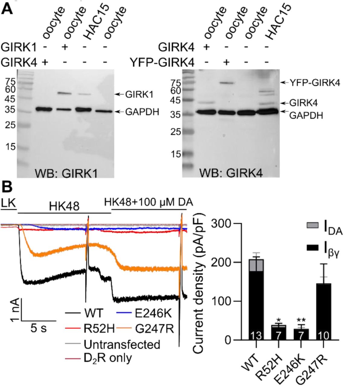
Figure Lengend Snippet: (A) Western blots of HAC15 cells with GAPDH antibody (both panels) and GIRK1 antibody (left) and GIRK4 antibody (right) show that HAC15 cells express GIRK1 and GIRK4 channels. Oocytes expressing GIRK1, GIRK4, and YFP-GIRK4 were used as positive controls, naïve oocytes as negative control, and GAPDH as loading control. (B) Examples (left) and summary (right) of currents in HAC15 cells expressing GFP-GIRK1/GIRK4 WT , GFP-GIRK1/GIRK4 R52H , GFP-GIRK1/GIRK4 E246K , and GFP-GIRK1/GIRK4 G247R . Channels were expressed with D 2 dopamine receptor and Gβγ (I βγ ), thus the small I DA .
Article Snippet: Protein samples (35µg) were electrophoresed on 12% polyacrylamide-SDS gel and transferred to nitrocellulose membranes for Western blotting with antibodies; anti-GIRK1 at 1:300 dilution (Alomone Labs, APC-005) or
Techniques: Western Blot, Expressing, Negative Control

Journal: bioRxiv
Article Title: A revised mechanism of action of hyperaldosteronism-linked mutations in cytosolic domains of GIRK4 (KCNJ5)
doi: 10.1101/866202
Figure Lengend Snippet: Oocytes expressed GIRK4 mutants and GIRK4 WT , with or without Gβγ. Cells were exposed to 4 concentrations of VU0529331 (1, 5, 20, and 60 μM). Only GIRK4 WT and GIRK4 G247R mutant responded to VU0529331. (A) I basal of GIRK4 WT and GIRK4 G247R . GIRK4 WT and GIRK4 G247R react similarly to VU0529331. (B) I βγ of GIRK4 WT and GIRK4 G247R . GIRK4 WT had larger current at 0 μM VU0529931, while GIRK4 G247R did not show any response to concentrations lower than 20 μM VU0529331. (C) Normalized I basal (I/I control ). GIRK4 WT and GIRK4 G247R were activated similarly by VU0529331. (D) I/I control of I βγ show that GIRK4 G247R was activated by VU0529331 stronger than GIRK4 WT , in relative terms. (E) I-V relationship of GIRK4 WT (top) and GIRK4 G247R (bottom) without Gβγ. (F) I-V relationship of GIRK4 WT (top) and GIRK4 G247R (bottom) with Gβγ.
Article Snippet: Protein samples (35µg) were electrophoresed on 12% polyacrylamide-SDS gel and transferred to nitrocellulose membranes for Western blotting with antibodies; anti-GIRK1 at 1:300 dilution (Alomone Labs, APC-005) or
Techniques: Mutagenesis
Journal: The Journal of Neuroscience
Article Title: Oxytocin-Modulated Ion Channel Ensemble Controls Depolarization, Integration and Burst Firing in CA2 Pyramidal Neurons
doi: 10.1523/JNEUROSCI.0921-22.2022
Figure Lengend Snippet: K ir channel subtype mRNA expression level in dHP CA2 region
Article Snippet: VU 0134992, VU 590 dihydrochloride,
Techniques: Expressing